
Field Note 1 · Protein design
Protein design is a network, not a pipeline
Backbone design, sequence design, evaluation: a familiar starting point for a field with many routes and rapidly evolving methods.
What does it mean to design a protein?
A protein’s amino-acid sequence helps determine its three-dimensional structure and what it can do. Protein design starts with a desired property or function and asks which sequences might realise it. That can mean adapting an existing protein or proposing one with a new sequence and fold. The objective might be a research reagent, a catalyst or a component for a biomaterial.
This reverses the familiar question of structure prediction. Prediction asks what structure a sequence may adopt. Design asks which sequence and structure could serve a purpose. The two are closely connected: predictions help assess proposals, while measurements reveal which parts of the design hypothesis hold up.
A familiar starting point
Protein design is often introduced as three tasks: propose a backbone, choose an amino-acid sequence and evaluate the resulting design. RFdiffusion, ProteinMPNN and AlphaFold2 illustrate these roles in a widely used structure-based approach.
- Backbone designRFdiffusion
Proposes the protein’s three-dimensional backbone.
- Sequence designProteinMPNN
Proposes amino-acid sequences for a given backbone.
- EvaluationAlphaFold2
Predicts a structure from sequence for structural assessment.
Different questions need different approaches
There is no single protein-design problem. Improving an existing protein starts with a sequence and often measurements of its behaviour. Designing a new fold starts with structural requirements. A binding project adds questions about the target and its biological context. An enzyme project asks whether a protein can support a chemical function, while an assembly project concerns how several components fit together.
These projects draw on different kinds of evidence. A sequence collection may reveal patterns in a protein family; a structure can expose geometry; biochemical measurements tell us how a molecule behaves under tested conditions. Language models, structural models and physicochemical descriptions provide different representations of this information. They can complement one another, but their outputs answer different questions.
The three-stage illustration therefore explains a useful set of roles. It does not define every project’s starting point or imply that one combination of tools is suitable for every scientific objective.
Why the field looks more like a network
The three tasks are useful landmarks, but individual tools can cover more than one of them. Some approaches start from an existing protein; others generate new structures or work directly with sequences. BindCraft, for example, integrates design and assessment in one pipeline. It belongs on the same map, but occupies a different role from a single backbone generator.
The scientific question also changes the route. A binder, an enzyme and a membrane protein present different challenges. Folding is one part of the problem; function, specificity, stability and the biological environment can matter just as much. Evidence that supports one property may say little about another.
The field is moving rapidly. New models, integrated approaches and experimental results keep expanding what can be attempted and changing how methods are assessed. The different approaches taken by RFdiffusion and BindCraft show how varied this landscape has become. A useful overview therefore needs to show the roles and relationships between methods, alongside the evidence behind them.
Explore the network
The interactive map accompanies this Field Note. Explore the stations, search for a method and follow its source links. On a phone, the searchable station list provides the same tool descriptions and references.
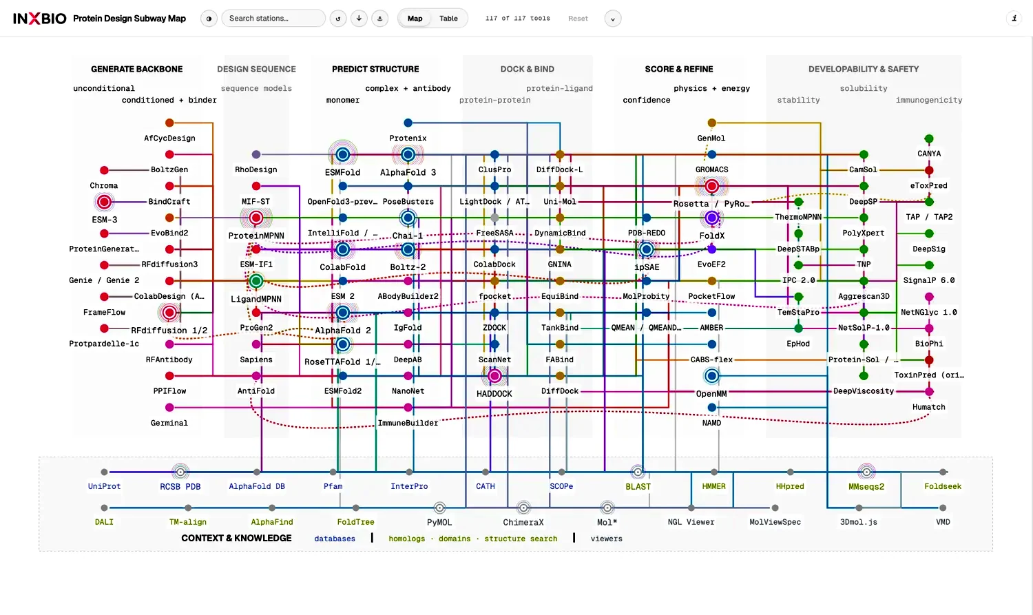
The map is adapted from Srikanth Lingappa’s Protein Design Subway Map, distributed under the MIT licence. InXBio’s additions include tool documentation, source links and the browser interface. This educational companion retains the upstream attribution.
How to read the map
Stations and lines. Each station represents a tool. Coloured lines group scientific aims, such as structure prediction, enzyme design or developability. They describe where a tool can contribute, rather than ranking its quality. Transfer stations mark tools used across several lines.
Regions and context. The main regions run from backbone and sequence design through structure prediction, docking, scoring and developability. Databases, structure search and viewers occupy a separate context band. A station’s region describes its role; its line colours show the kinds of work it supports.
Open a station. Select a tool to read its description and follow links to the original paper and software. Search helps you find a method without tracing every line. On phones, the station list offers the same descriptions and references.
Quality gates. These criteria help assess a result before the next step. Open a station or connection for task-specific thresholds and sources; the box below explains the main checks.
Quality gates: scores and checks
- Structural evaluation. Predicted structures are often a critical first step in evaluating a design’s fold and binding geometry. Tools such as AlphaFold, Boltz and ESMFold provide predicted coordinates; the appropriate model depends on whether a single protein or a complex is being assessed. 5
pLDDT, PAE and ipTM are confidence estimates supplied by prediction models. Fold RMSD is calculated by comparing structures, while ipSAE is computed from PAE. The available confidence scores vary by model.
- Binder pLDDT (predicted local distance difference test) measures confidence in the binder’s local structure. AlphaFold’s scale is 0–100: >90 very high confidence, 70–90 confident, 50–70 low and <50 very low. 6
- Fold RMSD (root-mean-square deviation) measures agreement between the designed and repredicted backbone after alignment. Range: 0 upwards, in ångströms (Å); lower is closer. <2 Å is a common self-consistency benchmark cutoff. 7
- Interface PAE (predicted aligned error) estimates uncertainty in the relative placement of residues across chains. Range: 0 upwards, in Å, with a model-specific upper limit; lower means greater confidence. Selection cutoffs depend on the task and model. 6
- ipTM (interface predicted template modelling score) measures confidence in the relative placement of interacting chains. Range: 0–1. AlphaFold-Multimer guidance: >0.8 confident, 0.6–0.8 uncertain, <0.6 low confidence. 6
- ipSAE (interaction prediction score from aligned errors) assesses interfaces using reliable interchain PAE values and adjusted length normalisation. Range: 0–1; higher indicates greater confidence. Its selection cutoff is benchmark-specific. 8
- Pose and interface quality. Pose recovery checks whether the intended binding orientation is reproduced. Clash checks identify overlapping atoms; interface chemistry examines packing, hydrogen bonds and unsatisfied polar groups.
- Developability. Assess whether the protein can be produced, remains stable and soluble, and resists aggregation under the intended conditions.
- Specificity evidence. Compare binding to the intended target with relevant alternatives. Experimental comparisons establish selectivity; structural confidence scores alone do not.
Connections are a guide to the landscape. Combining tools still depends on compatible inputs, assumptions and intended use. The map is a starting point for understanding a method, rather than a prescribed workflow or a ranking of tools.
What an evaluation can tell you
A predicted structure can support a design hypothesis. It does not establish binding, selectivity or thermal stability experimentally. Confidence scores and other computational assessments describe particular aspects of a model; their meaning depends on the method and the task. Experimental results provide the evidence needed to judge performance in the intended setting.
Even a well-folded protein may be difficult to produce, behave differently in a mixture or fail to perform its intended function. A membrane environment, a binding partner or the conditions of a measurement can change the interpretation. These are reasons to keep the biological question visible throughout a project, rather than reducing success to one model score.
Experimental feedback also changes what is known. Results from successful and unsuccessful candidates can reveal a limitation in the starting assumptions or motivate a different scientific question. This is why the broader discovery process is iterative, even when a computational illustration is drawn as a straight line.
Two tools may both return a structure or a score while using different training data, assumptions and definitions. Their outputs need interpretation in the context of the original method and its evaluation. The station references in the map provide a route to that context.
For the molecules themselves, continue with Antibody or minibinder. For the record behind a computational workflow, read Agents, servers and the rules between them.
Background: a brief history of protein design
Selected milestones, from early designed proteins through David Baker’s Rosetta work to deep learning and the 2024 Nobel Prize.
- 1988
Early de novo proteins
Lynne Regan and William DeGrado design and experimentally characterise a four-helix protein, showing how folding principles can guide the creation of a new protein. 9
- 1998
David Baker and Rosetta
David Baker’s group enters the CASP structure-prediction assessment with Rosetta. Initially developed for predicting structures, Rosetta later becomes a shared platform for protein modelling and design. 10
- 2003
Top7: a new protein fold
Brian Kuhlman and colleagues in Baker’s group use Rosetta to design Top7, a fold not previously observed in nature. Its experimentally determined structure closely matches the design. 11
- 2008–2010s
From folds to functions
Computational enzyme-design studies appear in 2008. Work across the field expands to binding proteins and self-assembling structures, bringing biological function into protein design. 12
- 2021
AlphaFold2: better structure prediction
Jumper and colleagues at DeepMind publish AlphaFold2. More accurate structure prediction strengthens the computational assessment of candidate designs. 3
- 2022
ProteinMPNN: learned sequence design
Dauparas and colleagues in Baker’s group introduce ProteinMPNN, which proposes amino-acid sequences compatible with a given protein backbone. 2
- 2023
RFdiffusion: generating new backbones
Watson, Juergens and colleagues in Baker’s group introduce RFdiffusion, a generative model derived from RoseTTAFold that creates new protein backbones. 1
- 2024
The Nobel Prize in Chemistry
David Baker receives half the prize for computational protein design. Demis Hassabis and John Jumper share the other half for protein structure prediction. 13
Sources and attribution
- Watson et al. De novo design of protein structure and function with RFdiffusion. Nature (2023).
- Dauparas et al. Robust deep learning–based protein sequence design using ProteinMPNN. Science (2022).
- Jumper et al. Highly accurate protein structure prediction with AlphaFold. Nature (2021).
- Pačesa et al. One-shot design of functional protein binders with BindCraft. Nature (2025).
- Official model documentation: AlphaFold and AlphaFold-Multimer (Google DeepMind), Boltz (Boltz contributors) and ESMFold (Meta FAIR).
- EMBL-EBI Training. AlphaFold confidence scores: pLDDT, PAE and ipTM in AlphaFold-Multimer.
- Jendrusch M, Korbel JO. Efficient protein structure generation with sparse denoising models. Nature Machine Intelligence (2025).
- Dunbrack RL Jr. Rēs ipSAE loquunt: What’s wrong with AlphaFold’s ipTM score and how to fix it. bioRxiv (preprint) (2025).
- Regan L, DeGrado WF. Characterization of a helical protein designed from first principles. Science (1988).
- Rosetta Commons. History of Rosetta. See also Nobel Prize Outreach, popular information for the 2024 Nobel Prize in Chemistry (2024).
- Kuhlman B et al.. Design of a novel globular protein fold with atomic-level accuracy. Science (2003).
- Huang, Boyken and Baker. The coming of age of de novo protein design. Nature (2016).
- The Royal Swedish Academy of Sciences. The Nobel Prize in Chemistry 2024. Press release, 9 October 2024.
- Yang et al. The past, present and future of de novo protein design. Nature (2026).
- Chu, Lu and Huang. Sparks of function by de novo protein design. Nature Biotechnology (2024).
- Kortemme. De novo protein design—From new structures to programmable functions. Cell (2024).
- Ingraham et al. Illuminating protein space with a programmable generative model. Nature (2023).
- Ahern et al. Atom-level enzyme active site scaffolding using RFdiffusion2. Nature Methods (2026; published online in 2025).