
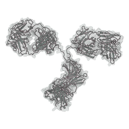
Antibody, IgG (immunoglobulin G)
About 150 kDa · about 1,300 amino acids, four chains
Two identical arms bind the antigen and the Fc stem recruits the immune system, which is also what secondary reagents and protein A columns recognise.
Field note 2
Antibodies and designed minibinders offer different sizes and binding surfaces. The useful comparison separates molecular format, discovery route and performance in the intended assay.
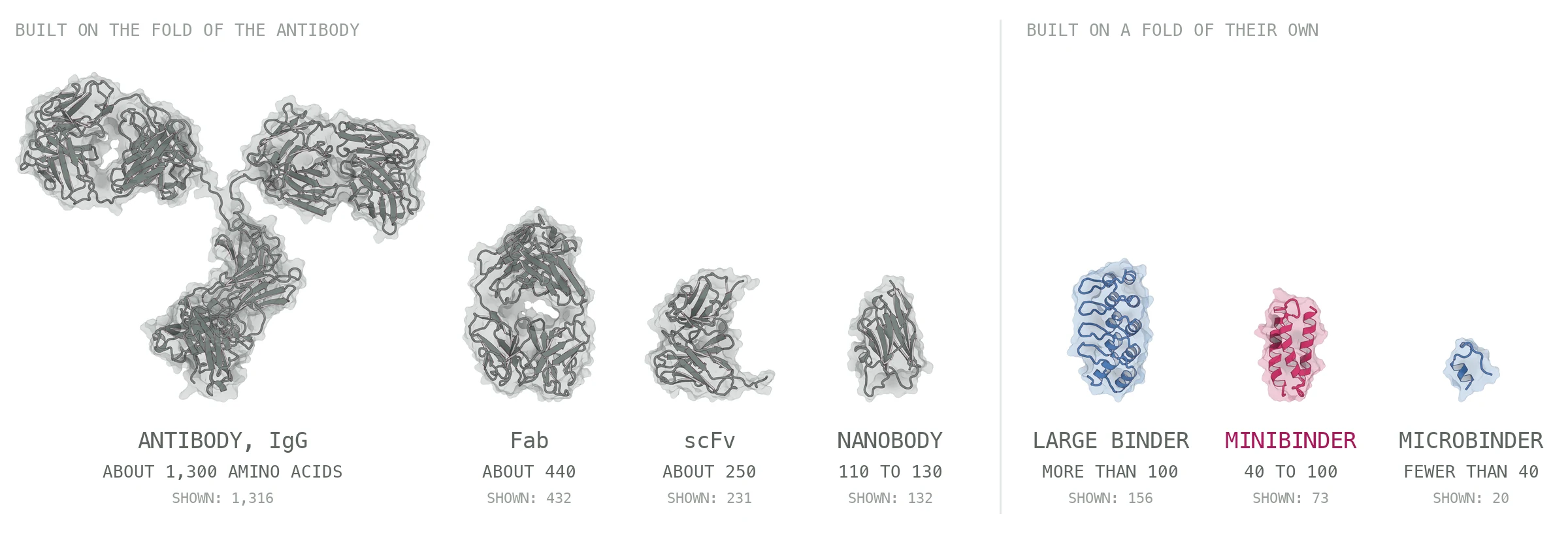
The convenient story is that antibodies come out of an animal and minibinders out of a computer, but it stopped being true some time ago: antibody fragments have been selected from phage libraries since 1990, 2 and since 2025 variable domains, single-chain fragments and whole antibodies can be designed against an epitope chosen in advance, with the designed loops confirmed by cryo-electron microscopy. 7 How a binder was obtained therefore says nothing about what it is.
The fold does. Every format on the left keeps the immunoglobulin fold, a sandwich of β-sheets whose paratope, the surface that contacts the target, is carried by the loops at one end. Every format on the right is built on a fold chosen for the task, and for small designed binders that fold is usually a bundle of three helices held together by buried hydrophobic side chains rather than by disulfide bonds.
Fold and size constrain the binding surface, but they do not by themselves determine which epitopes a binder can recognise. Antibody loops, single domains and designed protein surfaces span different geometries. Accessibility depends on the particular binder, target and biological state.
Built on the antibody fold Raised in an animal, selected from a library, or now designed
About 150 kDa · about 1,300 amino acids, four chains
Two identical arms bind the antigen and the Fc stem recruits the immune system, which is also what secondary reagents and protein A columns recognise.
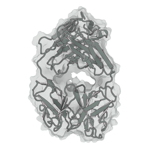
About 50 kDa · about 440 amino acids, two chains
One antigen-binding arm, without Fc-mediated effector functions. A Fab retains one binding site but loses the bivalent avidity of intact IgG. Removing Fc removes its Protein A binding site, although some VH3-family Fabs still bind Protein A through their variable domain. Structural evidence.
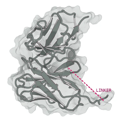
About 25 to 30 kDa · about 250 amino acids, one chain
A single-chain variable fragment: a flexible peptide linker joins the heavy- and light-chain variable domains into one chain, retaining all six antigen-binding loops. The rose dashed line is schematic; the linker is absent from the source antibody structure.
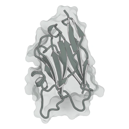
About 12 to 15 kDa · 110 to 130 amino acids, one domain
The variable domain of the heavy-chain antibodies that camelids make, with an unusually long CDR3 loop that can reach into a pocket a conventional paratope cannot enter.
Built on a fold of its own Computed against the structure of the target
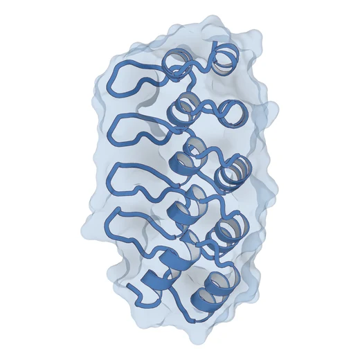
More than 100 amino acids
A designed protein above the minibinder range, with more surface available for contacts but more chain to express, fold and keep soluble.
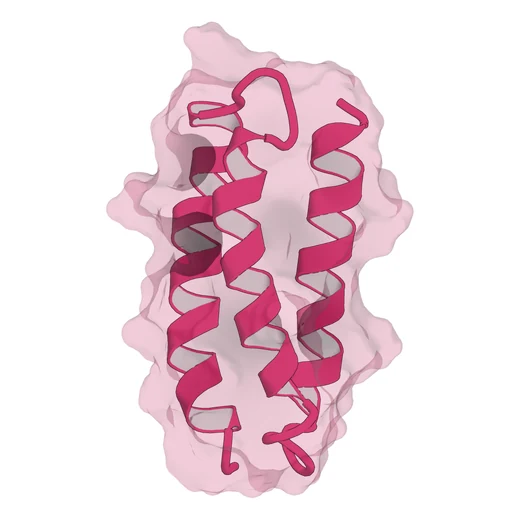
About 4 to 11 kDa · 40 to 100 amino acids, one chain
Often a compact helical fold. Some published miniproteins tolerate demanding conditions, but stability and solubility must be measured for each candidate.
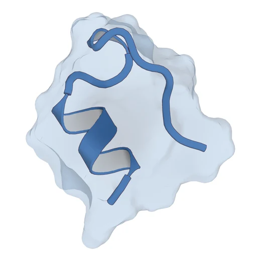
Fewer than 40 amino acids
A very small designed protein or peptide. Chemical synthesis may be practical, but binding surface, fold and experimental suitability depend on the molecule.
Here, 40 to 100 amino acids is a working convention for minibinder size, not a universal definition. Nanobodies and scFvs retain an immunoglobulin fold; a small designed scaffold is not established as a binder until its binding has been demonstrated.
Antibodies have had more than a century of method development behind them, designed binders about twenty years, and most of what matters in the second column arrived in the last five, once structure prediction became reliable enough to filter designs before anything was expressed.
The antibody fold
Folds of their own
Three computational steps make a binder today: a backbone is generated against the chosen epitope, a sequence is designed that folds into that backbone, and the complex is predicted again so that only designs the prediction confirms are ordered. Each step has several tools that will do the job, which is the subject of field note 1, and a review of 2026 sets out where the remaining failures are. 8
| Antibody | Designed minibinder | |
|---|---|---|
| Size | About 150 kDa. Roughly 1,300 amino acids in four chains. | About 4 to 11 kDa. 40 to 100 amino acids in one chain. |
| Fold | The immunoglobulin fold, binding done by loops. | Chosen for the target, usually a bundle of helices. |
| Binding surface | Loop-based surface; geometry varies by antibody format. | Compact surface; access depends on the designed geometry. |
| Where it comes from | An animal, a library, or, since 2025, design as well. | Designed against a structure or a model. |
| Choosing the site | Epitope choice can be guided by antigen presentation, selection or design. | Named before anything is designed. |
| Telling relatives apart | Specificity can be included in selection and tested experimentally. | Counter-targets can inform computational ranking; specificity is tested. |
| Measured affinity | Depends on the clone, optimisation and assay. | Established by experiment; a computational score is not an affinity measurement. |
| Stability | Depends on the format, sequence and conditions. | Some designed miniproteins tolerate heating; each candidate needs measurement. 5 |
| Where it wins | Abundant soluble antigens, established workflows, Western blot and fixed tissue. | Assays that benefit from a compact reagent with the required measured properties. |
Choose a format around the epitope, assay and required functions. Established IgG reagents remain useful where assay compatibility, Fc function or existing validation matters. A designed minibinder is another option when compact size and a defined structural hypothesis are valuable. Neither route removes the need to measure binding, specificity and performance in the intended experiment.